
Research article / Open Access
2022; 1(1): 116-121 . doi: 10.31488/jpp.104
Intracellular Reverse Transcription of COVID-19 mRNA Vaccine In vitro in Human Cell: Persistence Evaluation Needed
Luisetto M1, Naseer Almukthar2, Tarro Giulio3, Gamal abdul Hamid4, Farhan Ahmad Khan5 , Edbey K6 , Mashori Gulam Rasool7 , Nili behzad8, Fiazza C9, Cabianca L10 , ilnaf Ilman Student11, Yesvi A. Rafa12, Oleg yurevich13
1. Applied pharmacologist, professorship toxicology IMA academy, Italy 29121
2. Professor, Department of Physiology/College of Medicine, University of Babylon, Iraq
3. President of the T & L de Beaumont Bonelli Foundation for Cancer Research, Naples Italy
4. Professor Hematology Oncology, University of Aden, Yemen
5. Professor, Department of Pharmacology, Jawaharlal Nehru Medical College, AMU, Aligarh.
6. Professor, Department of Chemistry, Libya Physical Chemistry, University of Benghazi, Libya
7. Professor, Department of Medical & Health Sciences for Woman, Peoples University of Medical and Health Sciences for Women, Pakistan
8. Ahmadabadi Nano Drug Delivery, (a Product Development Firm), United States
9. Pharmacologist, Independent researcher Italy 29121 PC area
10. Biomedical laboratory Turin Italy Citta’ della Salute
11. Dhaka Residential Model College, Dhaka, Bangladesh
12. University of Nebraska-Lincoln,NE , USA Majoring in Biological sciences -Human Health and Disease.
13. President IMA Marijnskaya academy Russia
*Corresponding author:Luisetto M Applied pharmacologist, professorship toxicology IMA academy, Italy 29121
Abstract
The In vitro profile of action of some mRNA covid vaccine not only to Produce translates for the production of the spike protein but also the intracellular reverse transcription in some human cells. In this study it was verified effect on different times: at 6- 24- 48 hours and the results are clear. Related to these aspects, it is needed to be verified during the time 3-6- 12 -24 hours and more time month what happen. This can be useful to complete the profile of security for spike protein molecules production inducers related to this aspect.
Keywords: COVID-19 mRNA vaccine, intracellular reverses transcription, human cells, in vitro, Toxicology, Molecular biology
Introduction
Related m-RNA vaccine it is relevant to observe that they were introduced in rapid way to fight covid-19 spread. This first received by health authority’s emergency approval, and then complete approval by FDA on December 18, 2020. The time under clinical evaluation was shorter then classic vaccine required.
The mechanism involved in these new products implies a transduction of the genetic code of the m-RNA used to produce SPIKE protein by human ribosome. But as showed by some In Vitro study by Alden M [1] is the first in vitro study on the effect of COVID-19 mRNA vaccine BNT162b2 on human liver cell line. The present evidence on fast entry of BNT162b2 into the cells and subsequent intracellular reverse transcription of BNT162b2 mRNA into DNA”. This research was performed at 6-24-48 hours and the result shows this effect during this period. But what happen during period more prolonged as 3-6-12-24 month or more?.
According to Kaján GL et al. [2] “Viruses have been infecting their host -cells since the dawn of life, and this extremely long-term coevolution gave rise to some surprising consequences for the entire tree of life. It is hypothesized that viruses might have contributed to the formation of the first cellular- life form or that even the eukaryotic cell- nucleus originates from an infection by a coated virus. The continuous struggle between viruses and their hosts to maintain at least a constant fitness level led to the development of an unceasing arms race, where weapons are often shuttled between the participants.
It is generally accepted that an RNA-based world preceded today’s DNA- and protein-based one. RNA- viruses and especially the capsid less viroid might resemble this ancient world. The evolution of viruses—which has been especially rapid for RNA viruses—destroyed the possible signal of genetic relatedness a long time ago, making phylo-genetic reconstruction already impossible. Endogenous viral elements (EVEs) are viral genes or sometimes complete genomes inserted into host -genomes. As genome integration is a compulsory step in retroviral replication, most of these elements are of retroviral origin, but further virus families were detected as well: hepa-dna-viruses, adeno-associated viruses, herpes-viruses and others. If the insertion occurs in the germ cell line, the inserted sequence stretch might get inherited. And if it provides a selective advantage, e.g. protection from a viral infection, the genomic change will be fixed in the population. In case this advantage disappears, the inserted stretch might mutate over time and lose its protective nature”.
A virocentric perspective on the evolution of life Koonin EV et al. “Although viruses extensively exchange genes with their hosts, there exists a set of viral hallmark -genes that are shared by extremely diverse groups of viruses to the exclusion of cellular life forms. Coevolution of viruses and host defense systems is a key aspect in the evolution of both viruses and cells, and viral genes are often recruited for cellular functions. Together with the fundamental inevitability of the emergence of genomic parasites in any evolving replicator system, these multiple lines of evidences reveal the central role of viruses in the entire evolution of life. Viruses as nature’s genomic laboratory.
All cellular- life forms employ a single, strictly defined strategy for genome replication and expression whereby double-stranded (ds) DNA is the replicating form of the genome that is transcribed into single-stranded (ss) RNA some of which is then translated to produce proteins. The only variation on this theme is occasional reverse transcription of RNA that is typically non-essential for genome replication, an important exception being the telomere synthesis in eukaryotes. In a sharp contrast, viruses exploit effectively all possible combinations of DNA and RNA interconversions: the replicating genome that is incorporated into virions can be represented by either RNA or DNA, and the replication-expression cycles of RNA viruses can include a DNA- intermediate and vice versa The key observation on the viral hallmark-genes is that they possess only distant homologs in cellular life forms (whenever close homologs of these genes are detected, they appear to be of viral origin) and yet form a network that connects almost the entire virus- world).
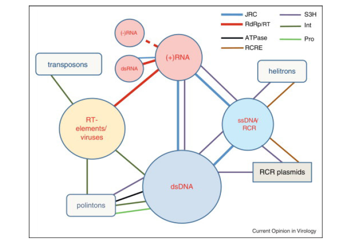
Figure 1:The network of hallmark- genes connecting different classes of viruses and capsid-less selfish elements. The colored -circles show classes of viruses and related capsid-less selfish elements and other shapes show distinct classes of non-viral selfish elements. The size of each shape roughly reflects the abundance and diversity of the respective class. The color-coded edges connecting the shapes denote shared hallmark- genes; the thickness of each line roughly reflects the prevalence of the respective gene in the corresponding classes of viruses and selfish- elements (in most cases, any given hallmark gene is present only in a subset of the class members). The dashed line reflects the tentative link between RNA-dependent RNA polymerases of positive-strand and negative-strand RNA viruses. JRC, Jelly Roll Capsid protein; RdRp, RNA-dependent RNA polymerase; RT, reverse- transcriptase; S3H, superfamily 3 helicase; Int, integrase; ATPase, packaging ATPase of the FtsK family; Pro, C5-family thiol protease; RCRE, rolling circle replication (initiation) endo-nuclease. Compared with the original list of viral hallmark genes, integrase and thiol proteases were additionally included whereas several genes that are widespread among diverse dsDNA viruses but not found in other classes of selfish elements are not shown. The Int gene is present in numerous but not all prokaryotic and eukaryotic DNA- transposons. Helitron’s are eukaryotic transposons that replicate via the RCR mechanism. Polinton’s are large, self-replicating eukaryotic transposons [3].
The parsimonious explanation of these findings appears to be that the hallmark- genes became isolated from the cellular genomes at the earliest stages of evolution and ever since comprised the framework of the temporally and spatially continuous, expanding virus world B (Figure 1).
Materials and Methods
With an observational point of view some relevant literatures for the scope of this work is reported. All articles came from scientific data base. An experimental hypothesis of work is submitted. After this process a global conclusion will be provided.
According to Walter Doerfler et al. [4] “Life has developed in a DNA, most likely preceded by RNA, world. We all live in this DNA world starting in our backyards when we dig the earth with uncounted microorganisms and their DNA buried in age-old debris of decaying animal and plant remnants. Just consider the annually shed foliage. With our daily food supply, we take up foreign DNA in huge quantities that is not momentarily digested to mononucleotides but transiently persists in the form of fragments that are capable of entering the human organism and becoming distributed in different human organ systems. DNA is a very stable molecule”
According to Forterre P et al. [5]. “The transition from the RNA to the DNA world was a major event in the history of life. The invention of DNA required the appearance of enzymatic activities for both synthesis of DNA precursors, retro-transcription of RNA templates and replication of single and double-stranded DNA molecules.”
The replication and Expression Strategies of Viruses by Rampersad S et al. [6] “Regardless of their genetic constitution, viral genomes are replicated, expressed, and assembled in association with living host cells. These entities do not undergo division, but rather generate new particles through the assembly of preformed components. Some viruses go further by modifying cellular metabolism to create a more favorable environment for viral gene expression. In a few instances, these clever strategies also facilitate escape from the host’s defense responses. The process of replication is typically divided into the phases of attachment, entry, uncoating, genome replication and expression, assembly, maturation, and finally, egress or release from the host cell. In this chapter, we address genome replication and expression, including the multiple strategies that serve to control gene expression and ensure preferential propagation of the virus.”
Origins and evolution of viruses of eukaryotes by Eugene V et al. [3] “According to the widely accepted RNA world hypothesis, the RNA-only replication cycle antedates reverse transcription and DNA-based replication (Bernhardt, Gilbert, Neveu et al. Robertson et al. [7-10].
Sébastien Desfarges et al. [11]. “By definition, RNA viruses are not able to integrate their genome into the host chromosome, as their genetic information resides in RNA molecules and not DNA. The only exception to this are retroviruses, which are characterized by the reverse transcription of their viral RNA genome into a linear double-stranded DNA molecule (viral DNA intermediate), and thus the substrate for subsequent viral genome integration into the host genome. For retroviruses, integration is a mandatory step for productive infection. Apart from retroviruses, the genome of other RNA viruses has been recently identified in the host genome”.
Results
According to report of By Aldén M Intracellular Reverse Transcription of Pfizer BioNTech COVID-19 mRNA Vaccine BNT162b2 In Vitro in Human Liver Cell Line [1] “RT-qPCR results showed that Huh7 cells treated with BNT162b2 had high levels of BNT162b2 mRNA relative to housekeeping genes at 6, 24, and 48 h (Figure 2, presented in logged 2−∆∆CT due to exceptionally high levels). The three BNT162b2 concentrations led to similar intracellular BNT162b2 mRNA levels at the different time points, except that the significant difference between 1.0 and 2.0 µg/mL was observed at 48 h. BNT162b2 mRNA levels were significantly decreased at 24 h compared to 6 h but increased again at 48 h” [1].
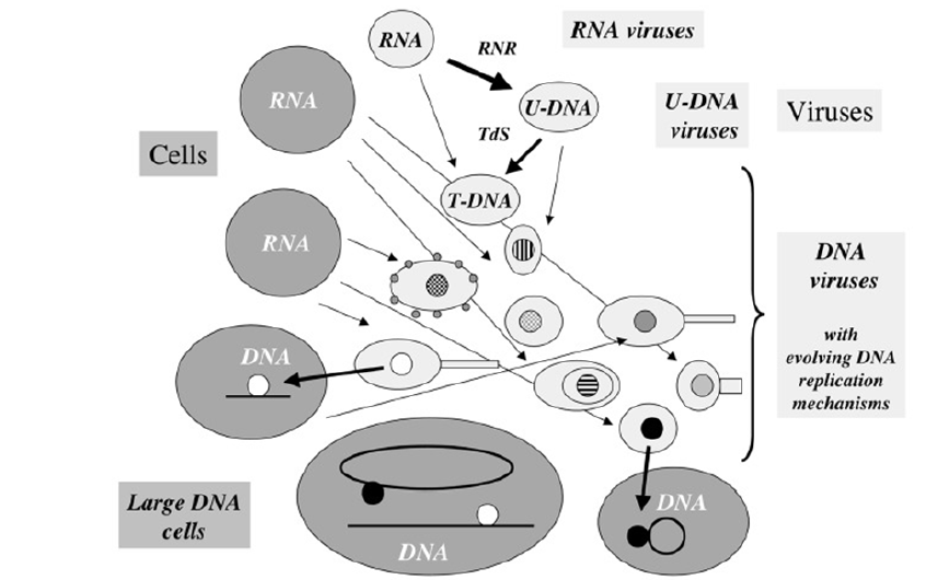
Figure 2:Evolution of DNA replication mechanisms in the viral world? This figure illustrates a coevolution scenario of cells and viruses in the transition from the RNA to the DNA world. Large gray circles or ovals indicate cells, whereas small light grey circles ovals (some with tails) indicate viruses. In this scenario, different replication mechanisms (inner circles with different colors) originated among various viral lineages after the invention of U-DNA and T-DNA by viruses (RNR= ribonucleotide reductase, TdS=Thymidylate synthase). These mechanisms evolved through the independent recruitment of cellular or viral enzymes involved in RNA replication or transcription (polymerases, helicases, nucleotide binding proteins) to produce enzymes involved in DNA replication (thin arrows). Two different DNA replication mechanisms (black and white circles) were finally transferred independently to cells (thick arrows). These two transfers can have occurred either before or after the LUCA. In the first case, the two systems might have been present in LUCA via cell fusion or successive transfers. One system could have also replaced the other in a particular cell lineage (from Origin and Evolution of DNA and DNA Replication Machineries The replication and Expression Strategies of Viruses by Sephra Rampersad and Paula Tennant [6]. “Regardless of their genetic constitution, viral genomes are replicated, expressed, and assembled in association with living host cells. These entities do not undergo division, but rather generate new particles through the assembly of preformed components. Some viruses go further by modifying cellular metabolism to create a more favorable environment for viral gene expression. In a few instances, these clever strategies also facilitate escape from the host’s defense responses. The process of replication is typically divided into the phases of attachment, entry, uncoating, genome replication and expression, assembly, maturation, and finally, egress or release from the host cell. In this chapter, we address genome replication and expression, including the multiple strategies that serve to control gene expression and ensure preferential propagation of the virus.”
We show here that SARS-CoV-2 RNA can be reverse-transcribed and integrated into the genome of the infected cell and be expressed as chimeric transcripts fusing viral with cellular sequences. Importantly, such chimeric transcripts are detected in patient-derived tissues. Our data suggest that, in some patient tissues, the majority of all viral transcripts are derived from integrated sequences. Our data provide an insight into the consequence of SARS-CoV-2 infections that may help to explain why patients can continue to produce viral RNA after recovery”. SARS–CoV–2 Spike Impairs DNA Damage Repair and Inhibits V (D) J Recombination In Vitro by Hui Jiang et al. [12,13].
Severe acute respiratory syndrome coronavirus 2 (SARS–CoV–2) has led to the coronavirus disease 2019 (COVID–19) pandemic, severely affecting public health and the global economy. Adaptive immunity plays a crucial role in fighting against SARS–CoV–2 infection and directly influences the clinical outcomes of patients. Clinical studies have indicated that patients with severe COVID–19 exhibit delayed and weak adaptive immune responses; however, the mechanism by which SARS–CoV–2 impedes adaptive immunity remains unclear. Here, by using an in vitro cell line, we report that the SARS–CoV–2 spike protein significantly inhibits DNA damage repair, which is required for effective V(D)J recombination in adaptive immunity. Mechanistically, we found that the spike protein localizes in the nucleus and inhibits DNA damage repair by impeding key DNA repair protein BRCA1 and 53BP1 recruitment to the damage site. Our findings reveal a potential molecular mechanism by which the spike protein might impede adaptive immunity and underscore the potential side effects of full-length spike-based vaccines” [13].
According to Proal AD et al. [14] “A growing number of studies show that some patients infected with SARS-CoV-2 do not successfully clear the virus over long periods of time [15-17]. In such studies, confirmation of SARS-CoV-2 in patient samples is generally assessed via identification of viral RNA and/or proteins. While identification of SARS-CoV-2 RNA could technically represent “inert” RNA, the possibility is unlikely because inert RNA in the human body is rapidly degraded [18].
According to panel A.Ciuffi “Retroviruses RNA virus, including the human immunodeficiency virus (HIV), are notorious for two essential steps of their viral replication: reverse transcription and integration. This latter property is considered to be essential for productive replication and ensures the stable long-term insertion of the viral genome sequence in the host chromatin, thereby leading to the life-long association of the virus with the infected cell. Using HIV as a prototypic example, the present review aims to provide an overview of how and where integration occurs, as well as presenting general consequences for both the virus and the infected host” [19].
Katzourakis A et al. “Integration into the nuclear genome of germ line cells can lead to vertical inheritance of retroviral genes as host alleles. For other viruses, germ line integration has only rarely been documented. Nonetheless, we identified endogenous viral elements (EVEs) derived from ten non-retroviral families by systematic in silico screening of animal genomes, including the first endogenous representatives of double-stranded RNA, reverse-transcribing DNA, and segmented RNA viruses, and the first endogenous DNA viruses in mammalian genomes “[20].
Anastasiya K et al. “Viruses hijack the host cell to replicate their RNA or DNA genomes and create progeny virions. An extreme form of viral parasitism is the integration of a viral genome DNA copy into the host cell DNA [22-23]. Recent studies reported a high frequency of reverse transcription and integration of severe acute respiratory syndrome coronavirus 2 (SARS-CoV-2) RNA in infected cells [12, 24].
High frequency of somatic integration of the RNA virus severe acute respiratory syndrome coronavirus 2 (SARS-CoV-2) into the DNA of infected cells was recently suggested, based on a number of observations. One key observation was the presence of chimeric RNA-sequencing (RNA-seq) reads between SARS-CoV-2 RNA and RNA transcribed from human host DNA” [21]. According to M. Pistello et al. [25] “Until a few years ago, integration was considered a must only for retroviruses.
More recently and, to our opinion, quite unexpectedly, it has been shown that integration takes place for many viruses, even those RNA viruses that entirely replicate in the cytoplasm and do not convert their RNA genome into DNA. The scientific community is struggling to find a mechanistic model that may explain how integration occurs for, for instance, bornaviruses, filoviruses, flaviviruses, picornaviruses and rabdoviruses, which have single-stranded RNA (either negative or positive polarity) genomes and whose replication is entirely cytoplasmic [26]. Less surprising, considering their nuclear replication, but nonetheless difficult to explain, is the case of the members of the Orthomixoviridae, whose segmented single-stranded negative polarity RNA genome, has been found integrated in insect cells [27]. The same consideration also applies to various single-stranded DNA viruses (circoviruses and parvoviruses) [26,27]. According to various molecular clocks, integration occurred several million years ago and, as happened for endogenized retroviruses, the viral genomes underwent gross rearrangement with conspicuous loss of genetic material.
Analyses beyond more pinpointing of integration sites and genome sequencing reinforced the idea that integration bears more advantages for the cell than the virus. As a likely result of host pressure, some endogenous retroviruses were eventually lost with a decline rate that differs among mammalian lineage and is particularly fast in humans [28].
Discussion and Conclusion
It is confirmed by published research the IN VITRO effect of some mRNA covid vaccine related intracellular reverse transcription in human liver cell line. Because this research provides informations in a window time of only 6-48 h it is relevant to observe also the pattern of persistence during more time of this activity (3-6-12-24-48 Month and more). This make possible to complete the safety profile of spike protein inducers by mRNA of exogen origin. The same according the author of this editorial it is needed to verify on field the complex toxicological profile during and effective time of 5-10 years and to deeply investigate the entity in quantitative methods of the transcription activity during the time.
Conflict of Interests
No
References
1. Aldén M, Olofsson Falla F, Yang D, et al. Intracellular Reverse Transcription of Pfizer BioNTech COVID-19 mRNA Vaccine BNT162b2 In Vitro in Human Liver Cell Line. Curr Issues Mol Biol. 2022; 44: 1115-1126. https://doi.org/10.3390/cimb44030073.
2. Kaján GL, Doszpoly A, Tarján ZL, et al. Virus-Host Coevolution with a Focus on Animal and Human DNA Viruses. J Mol Evol. 2020;88(1):41-56. doi:10.1007/s00239-019-09913-4.
3. Koonin EV, Dolja VV, Krupovic M. Origins and evolution of viruses of eukaryotes: The ultimate modularity. Virol. 2015; 479–480. https://doi.org/10.1016/j.virol.2015.02.039.
4. Doerfler W. Adenoviral Vector DNA- and SARS-CoV-2 mRNA-Based Covid-19 Vaccines: Possible Integration into the Human Genome - Are Adenoviral Genes Expressed in Vector-based Vaccines? Virus Res. 2021;302:198466.
5. Forterre P, Filée J, Myllykallio H. Origin and Evolution of DNA and DNA Replication Machineries. In: Madame Curie Bioscience Database [Internet]. Austin (TX): Landes Biosci. 2000-2013.
6. Rampersad S, Tennant P. Replication and expression strategies of viruses Viruses Mol. Biol. Host Interact. Appl Biotechnol. 2018; 55-82.
7. HS Bernhardt. The RNA world hypothesis: the worst theory of the early evolution of life (except for all the others)(a) Biol. Direct. 2012; 23.
8. W Gilbert. The RNA world. Nature. 1986; 319: 618.
9. M Neveu, HJ Kim, SA Benner. The “strong” RNA world hypothesis: fifty years old Astrobiology. 2013; 13 (4):391-403.
10. MP Robertson, GF Joyce. The origins of the RNA world Cold Spring Harb. Perspect Biol. 2012; 4: 5.
11. Desfarges S, Ciuffi A. Viral Integration and Consequences on Host Gene Expression. Viruses: Essential Agents of Life. 2012;147-175.
12. Zhang L, Richards A, Barrasa MI, et al. Reverse-transcribed SARS-CoV-2 RNA can integrate into the genome of cultured human cells and can be expressed in patient-derived tissues. Proc Natl Acad Sci U S A. 2021;118(21).
13. Jiang H, Ya-Fang Mei. SARS–CoV–2 Spike Impairs DNA Damage Repair and Inhibits V(D)J Recombination In Vitro Viruses. 2021; 13(10): 2056.
14. Proal AD, VanElzakker MB. Long COVID or Post-acute Sequelae of COVID-19 (PASC): An Overview of Biological Factors That May Contribute to Persistent Symptoms. Front Microbiol. 2021;12:698169. doi: 10.3389/fmicb.2021.698169. PMID: 34248921; PMCID: PMC8260991.
15. Liotti FM, Menchinelli G, Marchetti S, et al. Assessment of SARS-CoV-2 RNA test results among patients who recovered from COVID-19 with prior negative results. JAMA Intern Med. 2020;181:702–704. 10.1001/jamainternmed.2020.7570.
16. Sun J, Xiao J, Sun R, et al. Prolonged persistence of SARS-CoV-2 RNA in body fluids. Emerg Infect Dis. 2020; 26: 1834–1838. 10.3201/eid2608.201097.
17. Vibholm LK, Nielsen SS, Pahus MH, et al. SARS-CoV-2 persistence is associated with antigen-specific CD8 T-cell responses. EBioMedicine. 2021; 64:103230.
18. Fabre AL, Colotte M, Luis A, et al. An efficient method for long-term room temperature storage of RNA. Eur J Hum Genet. 2014; 22: 379–385.
19. A Ciuffi. The benefits of integration, Clinical Microbiology and Infection. 2016; 22: 324-332.
20. Katzourakis A, Gifford RJ. Endogenous Viral Elements in Animal Genomes. PLoS Genet. 2010; 6(11): e1001191. https://doi.org/10.1371/journal.pgen.1001191.
21. Anastasiya K, George K. SARS-CoV-2-Host Chimeric RNA-Sequencing Reads Do Not Necessarily Arise From Virus Integration Into the Host DNA. Frontiers Microbiol. 2021; 12.
22. Burns KH, Boeke JD. Human transposon tectonics. Cell 149, 740–752. doi: 10.1016/j.cell.2012.04.019.
23. Feschotte C, Gilbert C. Endogenous viruses: insights into viral evolution and impact on host biology. Nat Rev Genet. 2012; 13, 283–296. doi: 10.1038/nrg3199.
24. Ying Y, Xiao-Zhao L, Ximiao H, et al. Exogenous coronavirus interacts with endogenous retrotransposon in human cells. Front Cell Infect Microbiol. 2021; 11:609160. doi: 10.3389/fcimb.2021.609160.
25. M Pistello, G Antonelli. Integration of the viral genome into the host cell genome: a double-edged sword. Clin Microbiol Infect. 2016; 22: 296–298; DOI:https://doi.org/10.1016/j.cmi.2016.01.022.
26. Holmes EC. The evolution of endogenous viral elements. Cell Host Microbe. 2011; 10: 368-377.
27. Aiewsakun P, Katzourakis A. Endogenous viruses: connecting recent and ancient viral evolution. Virology. 2015; 479–80: 26-37.
28. Naville M. Warren IA, Haftek-Terreau Z, et al. Not so bad after all: retroviruses and LTR retrotransposons as a source of new genes in vertebrates. CMI. 2016; 22: 312-323.
Received: January 15, 2021;
Accepted: February 14, 2022;
Published: February 16, 2022.
To cite this article : Luisetto M, Almukthar N, Giulio T, et al. Plasma Rich in Growth Factors Treament for Patients with Refractary Endometrium Undergoing Embryo Criotransfer. Phase Iii Trial Protocol. Pharmacology and pharmacogenomics. 2022; 1(1): 116-121. doi: 10.31488/jpp.104.
© 2022 Luisetto M, et al.